Home>>Lyso-Tracker Green

Lyso-Tracker Green Sale

目录号 : GC20101

Lyso-Tracker Green是一种溶酶体(lysosome)绿色荧光探针,能通透细胞膜,可以用于活细胞溶酶体特异性荧光染色。

Lyso-Tracker Green Chemical Structure

规格 价格 库存 购买数量
50μl
¥707.00
现货

电话:400-920-5774 Email: sales@glpbio.cn

Customer Reviews

Based on customer reviews.

Sample solution is provided at 25 µL, 10mM.

产品文档

Quality Control & SDS

View current batch:

产品描述

Lyso-Tracker Green是一种溶酶体(lysosome)绿色荧光探针,能通透细胞膜,可以用于活细胞溶酶体特异性荧光染色。

Lyso-Tracker Green为采用Molecular Probes公司的DND-26进行了荧光标记的带有弱碱性的荧光探针,其中仅弱碱可部分提供质子,以维持pH在中性,可以选择性地滞留在偏酸性的溶酶体中,从而实现对于溶酶体的特异性荧光标记。中性红(Neutral Red)和吖啶橙(Acridine Orange)也都可以对溶酶体进行荧光染色,但中性红和吖啶橙的染色缺乏特异性。Lyso-Tracker Green适用于活细胞染色,但不适合用于固定后细胞的染色。

Lyso-Tracker Green的分子式为C18H26BClF2N4O,分子量为398.7,其最大激发光波长为504nm,最大发射光波长为511nm。Lyso-Tracker Green的化学结构式和激发、发射光谱图参考图1。


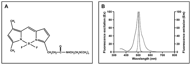
图1. Lyso-Tracker Green的化学结构式(A)和激发、发射光谱图(B)。

Lyso-Tracker Green是嗜酸性荧光探针,用于活细胞内酸性细胞器的标记和示踪。这些探针具有几个重要特征,包括高度选择靶向酸性细胞器和在纳摩尔浓度有效标记活细胞。Lyso-Tracker Green必须在极低浓度(通常约50nM)下才能获得优异的选择性。这些探针的滞留(retention)机制虽然没有被研究清楚,但很可能与酸性细胞器的质子化和滞留性有关,Lyso-Tracker Green探针的内吞作用动力学研究显示染料进入活细胞的摄入时间仅几秒即可。然而,这些溶酶体探针会导致溶酶体被碱化,长期孵育会诱使溶酶体pH值的增加。因此,建议成像前用探针孵育细胞的时间不能太久。Lyso-Tracker Green探针具体使用浓度和孵育时间需要根据自身实验条件和具体细胞种类来进行摸索以达到满意的染色效果。Lyso-Tracker Green染色活细胞溶酶体的效果请参考图2。

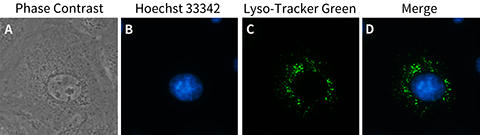
图2. Lyso-Tracker Green染色NRK-52E细胞(大鼠肾小管上皮细胞)的效果图。Hoechst 33342染色的NRK-52E细胞其细胞核呈现蓝色荧光;Lyso-Tracker Green染色的NRK-52E细胞其溶酶体呈现绿色荧光。

按照1:20,000的比例稀释,可以配制1000ml Lyso-Tracker Green工作液。

Chemical Properties

Cas No. SDF
分子式 C18H26BClF2N4O 分子量 398.7
溶解度 储存条件 Store at -20℃,protect form light
General tips 请根据产品在不同溶剂中的溶解度选择合适的溶剂配制储备液;一旦配成溶液,请分装保存,避免反复冻融造成的产品失效。
储备液的保存方式和期限:-80°C 储存时,请在 6 个月内使用,-20°C 储存时,请在 1 个月内使用。
为了提高溶解度,请将管子加热至37℃,然后在超声波浴中震荡一段时间。
Shipping Condition 评估样品解决方案:配备蓝冰进行发货。所有其他可用尺寸:配备RT,或根据请求配备蓝冰。

溶解性数据

制备储备液
1 mg 5 mg 10 mg
1 mM 2.5082 mL 12.5408 mL 25.0815 mL
5 mM 0.5016 mL 2.5082 mL 5.0163 mL
10 mM 0.2508 mL 1.2541 mL 2.5082 mL
  • 摩尔浓度计算器

  • 稀释计算器

  • 分子量计算器

质量
=
浓度
x
体积
x
分子量


*在配置溶液时,请务必参考产品标签上、MSDS / COA(可在Glpbio的产品页面获得)批次特异的分子量使用本工具。

计算

动物体内配方计算器 (澄清溶液)

第一步:请输入基本实验信息(考虑到实验过程中的损耗,建议多配一只动物的药量)
给药剂量 mg/kg 动物平均体重 g 每只动物给药体积 ul 动物数量
第二步:请输入动物体内配方组成(配方适用于不溶于水的药物;不同批次药物配方比例不同,请联系GLPBIO为您提供正确的澄清溶液配方)
% DMSO % % Tween 80 % saline
计算重置

Research Update

A series of meso amide BODIPY based lysosome-targeting fluorescent probe with high photostability and sensitivity

Anal Chim Acta 2022 May 1;1205:339771.PMID:35414392DOI:10.1016/j.aca.2022.339771.

Lysosomes are important organelles in physiological and pathological processes. It is of great significance to understand the mechanism of lysosome and monitor its movement and action at cellular level. Traditional lysosome trackers include Lyso-Tracker Green and Lyso-Tracker Red. However, both of them are tend to be photobleached easily and affected by pH variation, which is not conducive for long-term and real-time tracing of lysosomes in changeable environment. Herein, we designed a series of meso amide BODIPY based lysosome-targeting fluorescent probes. It was discovered that introduction of methyl group on amide is able to change the fluorescence characteristics of meso amide BODIPY. Among BODIPYs developed, Lyso-Me-1 exhibited outstanding lysosome-targeting ability in comparison with Lyso-Tracker Green confirmed by confocal microscope colocalization experiment. Moreover, continuous scanning of confocal microscope demonstrated that Lyso-Me-1 displayed improved photostability compared with Lyso-Tracker Green and Lyso-Tracker Red.

Role of biomimetic nanomaterials made from glioma cell- derived extracellular vesicles in targeted delivery of STAT3-siRNA

Zhong Nan Da Xue Xue Bao Yi Xue Ban 2022 Dec 28;47(12):1646-1654.PMID:36748374DOI:10.11817/j.issn.1672-7347.2022.210764.

Objectives: Glioma is the most common primary intracranial tumor and there is still no ideal treatment at present. Gene therapy, as one of the new methods for treating glioma, has attracted attention in recent years. But its application in treating glioma is very limited due to lack of effective delivery vectors. This study aims to investigate the feasibility of biomimetic nanomaterials made from glioma cells-derived extracellular vesicles (EV) for targeted delivery of signal transducers and activators of transcription 3 (STAT3)-small interfering RNA (siRNA) in treating glioma. Methods: First, U251 glioma cells-derived extracellular vessel (EVU251) was extracted by ultra-centrifugal method. Nanoparticle tracking analysis was used to characterize the particle size distribution, the transmission electron microscope was used to analyze the morphology, and Western blotting was used to verify the expression of srface characteristic protein. The homing ability was verified by cell uptake assay after labeling EVU251 with membrane dye kit PKH67; the EVU251 contents were removed by a low permeability method and then EVMU251 was prepared through a microporous membrane. Finally, the biomimetic nanomaterials EVMU251@STAT3-siRNA were prepared by loading STAT3-SiRNA with electro-dyeing method. The real-time quantitative PCR was used to quantify the successful encapsulation of siRNA, and the encapsulation and drug loading rate was calculated; then Cy5-labeled siRNA was used to evaluate the ability of biomimetic nanomaterials (EVMU251@CY5-siRNA) to target U251 cells. Lysosomal escape ability of the biomimetic nanomaterial was evaluated by lysosomal dye Lyso-Tracker Green. At last, the ability of EVMU251@STAT3-siRNA to knock down STAT3 gene and selective killing of U251 cells was detected by cell experiments in vitro. Results: The size of EVU251 ranged from 50 nm to 200 nm with a natural disc shape. The expression of extracellular vesicle marker proteins could be detected on the membrane of EVU251. The cell uptake assay demonstrated that it had homing ability to target U251 cells. After EVU251 was prepared as EVMU251@STAT3-siRNA, the particle size was (177.9±5.0) nm, the siRNA loading rate was (33.5±2.2)% and the drug loading rate was (3.24±0.21)%. The biomimetic nanomaterial EVMU251@STAT3-siRNA still had the ability to target U251 cells and successfully deliver siRNA to the cytoplasm without lysosomal degradation. The EVMU251@STAT3-siRNA can effectively knock down the expression of STAT3 gene and produce selective killing ability in U251 cells. Conclusions: The biomimetic nanomaterials EVMU251@STAT3-siRNA made from glioma U251 cells-derived extracellular vesicles can knock down STAT3 gene of U251 cells and produce selective killing effect, which can provide a new idea for the treatment of glioma.

Different heavy metals have various effects on Picea wilsonii pollen germination and tube growth

Plant Signal Behav 2015;10(4):e989015.PMID:25830714DOI:10.4161/15592324.2014.989015.

Heavy metal pollution has became one of the realistic matters of globality. Previous reports indicated that heavy metals could significantly inhibit pollen germination and tube growth. In the present study, comparative studies on the effects of different heavy metals (As, Hg, Cd, Cr and Cu) on in-vitro picea wilsonii pollen germination and tube growth were carried out. Microscopic evaluation revealed that different heavy metals had various degree of toxicity on P. wilsonii pollen tube development. As showed the most toxic effects on pollen germination, which was followed by Hg and Cd, while Cr and Cu showed relatively lower toxicity. Besides, pollen tubes showed varying shapes in response to different heavy metal stress. Pollen tubes treated with Cd, Hg and As were usually characterized by irregularly increasing diameters and swelling tips with distinct cytoplasimic vacuolation. On the other hand, except for the slightly increased diameters, no obvious abnormal shape were observed in tubes treated with Cr or Cu. Lyso-Tracker Green staining indicated that only Cd-treated pollen tubes showed numerous vacuole-like acidic organelles, though cytoplasmic vacuolization were also observed in pollen tubes treated with Hg and A. In brief, our data indicated that different heavy metals have various effects on Picea wilsonii pollen germination and tube growth, and that in-vitro pollen culture might be used as a competent system for biomonitoring of air pollution.